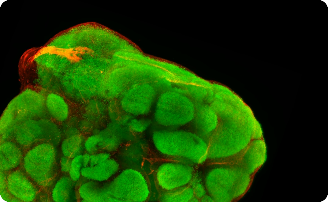
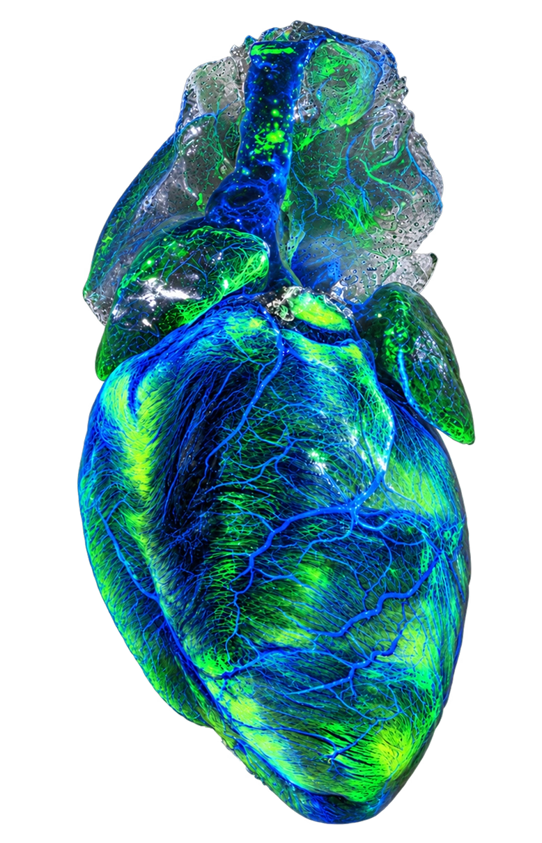
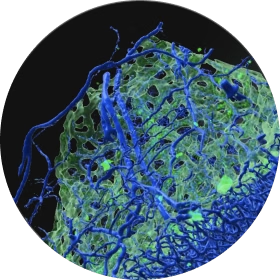
Colorful light-sheet scan of an organoid

Different labs have different needs. Explore the XL2 and UL systems

XL2 microscope and UL microscope system on a laboratory table

Supports multiple magnifications

Choose the right magnification for each specimen

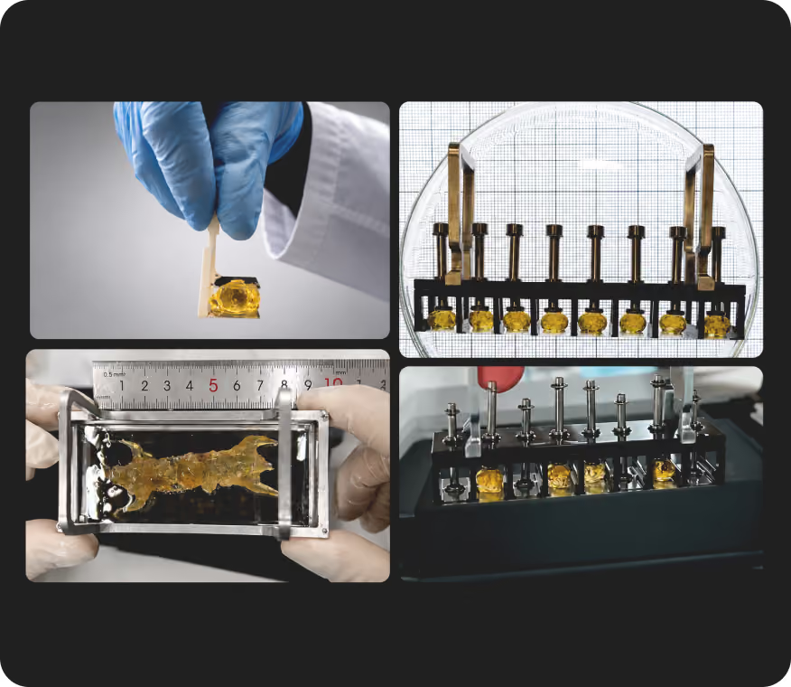
Flexible sample loading

Supports different sample sizes and holders

Smart objective & holder detection

Automatically detects installed objectives and sample holders

Integrated processing

Stitching, correction and 3D rendering in one workflow

High-resolution 3D imaging

Capture fine structures across cleared biological samples

Heated imaging chamber

Maintains stable temperature for aqueous imaging methods

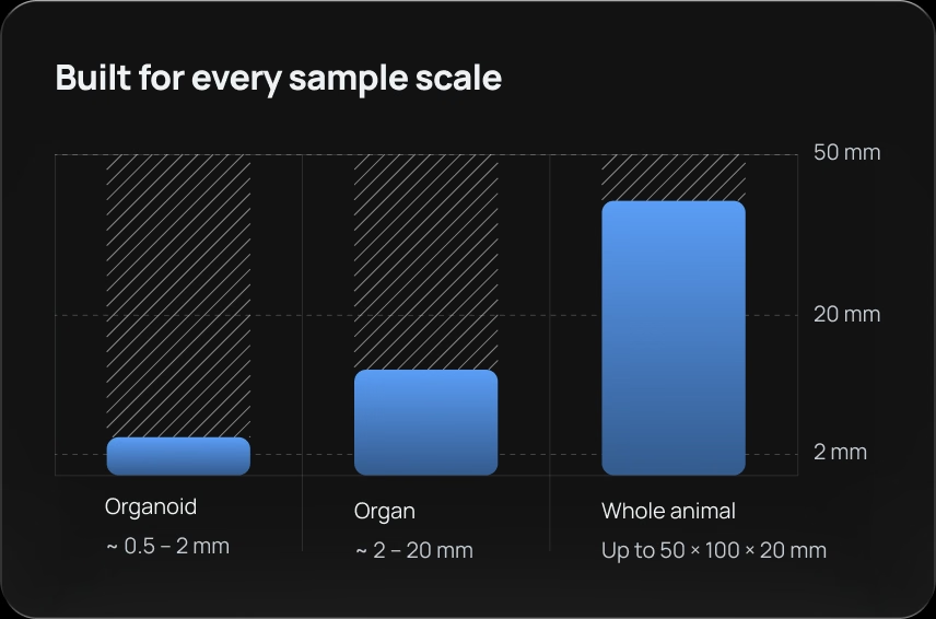
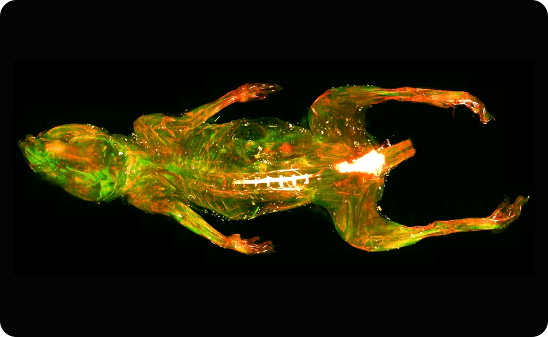
Built for every sample scale: organoid, organ and whole animal
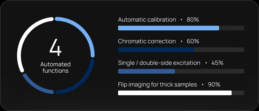
Four automated functions: calibration, chromatic correction, excitation and flip imaging
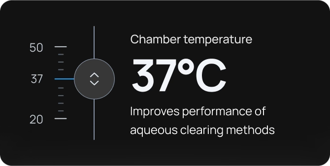
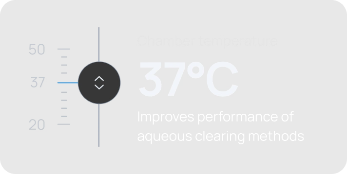
Chamber temperature 37°C. Improves performance of aqueous clearing methods.

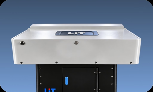
XL2 Lightsheet Microscope
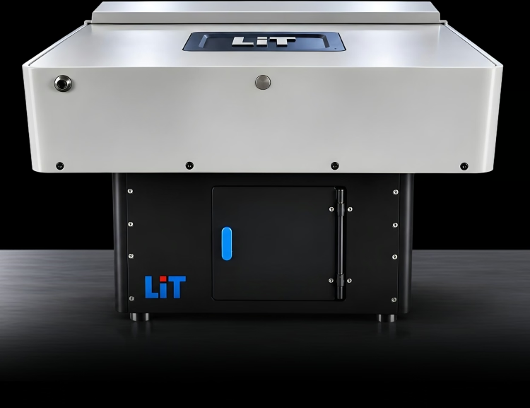
XL2 microscope scan

Supports multiple magnifications

Choose the right magnification for each specimen

Flexible sample loading

Supports different sample sizes and holders

Smart objective & holder detection

Automatically detects installed objectives and sample holders

Integrated processing

Stitching, correction and 3D rendering in one workflow

High-resolution 3D imaging

Capture fine structures across cleared biological samples

Heated imaging chamber

Maintains stable temperature for aqueous imaging methods

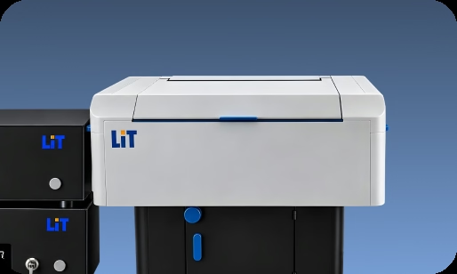
UL Lightsheet Microscope

UL microscope scan
Built for every sample scale: organoid, organ and whole animalFour automated functions: calibration, chromatic correction, excitation and flip imagingChamber temperature 37°C. Improves performance of aqueous clearing methods.
All scales

Built to answer the questions your science needs to answer

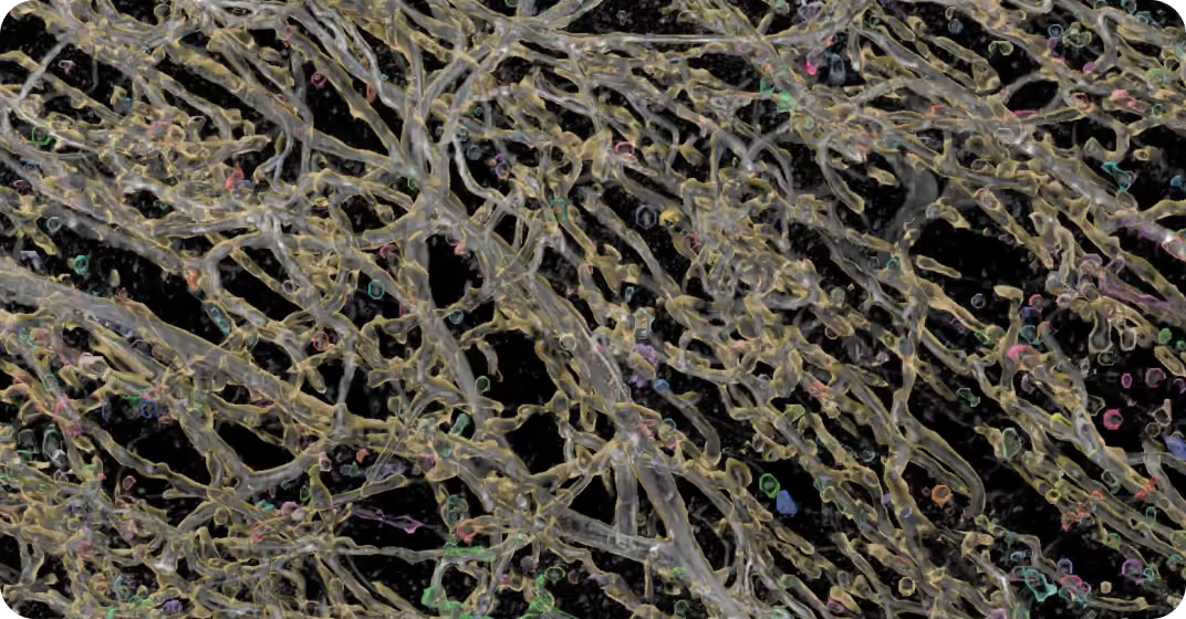
Microscopy visualization of neural tissue
Organoids

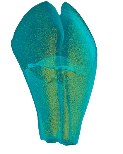
High-resolution imaging of 3D organoid models for structure, growth and phenotype analysis

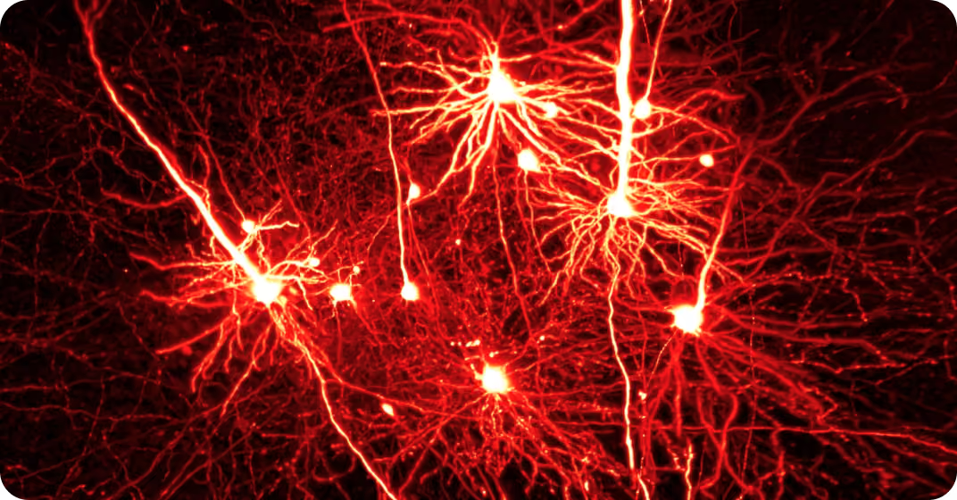
Microscopy visualization of neural tissue

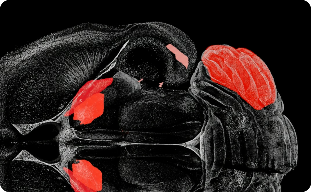
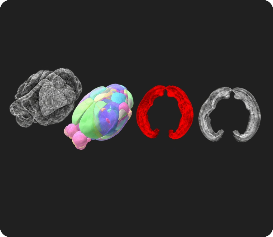
Whole-brain imaging for mapping, cell analysis, connectomics and vascular tracing across cleared neural tissue

Microscopy visualization of neural tissue

Large-volume imaging of cleared organs and tissues for structural, cellular and spatial analysis

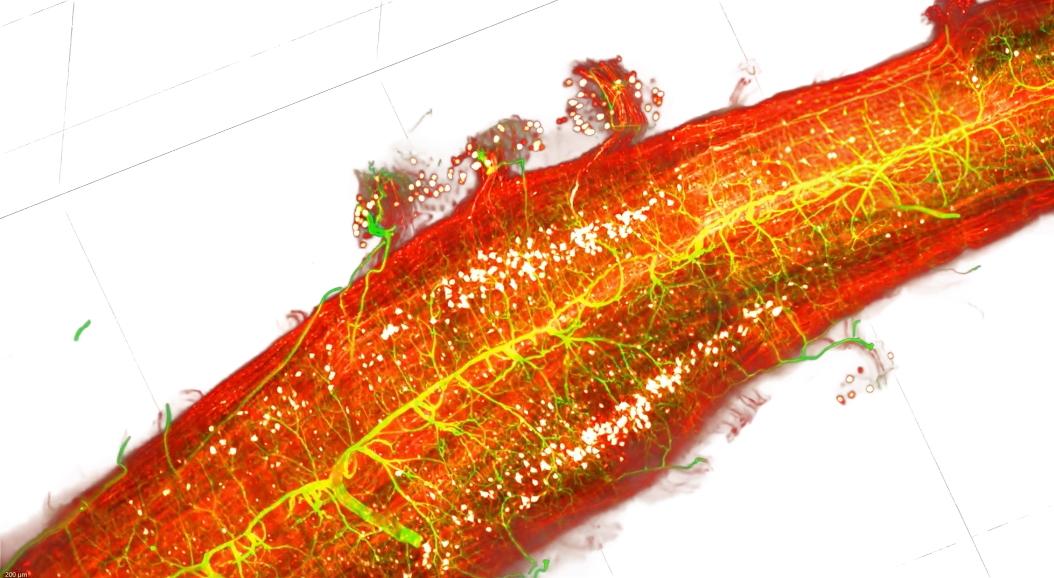
Microscopy visualization of neural tissue

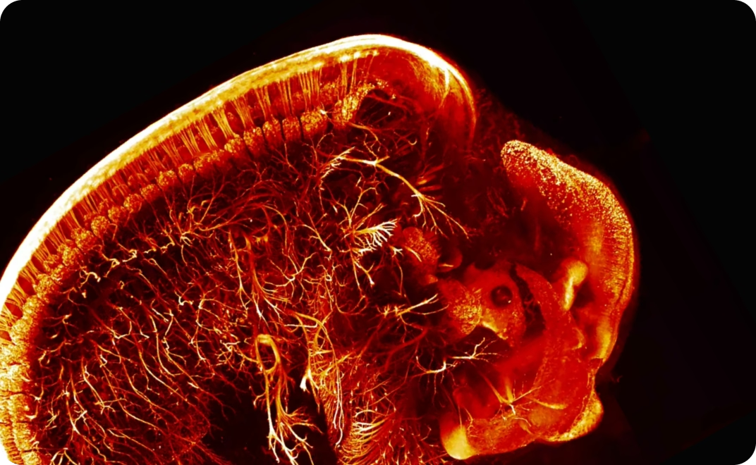
Visualize whole specimens across developmental stages to study morphology, growth and tissue organization

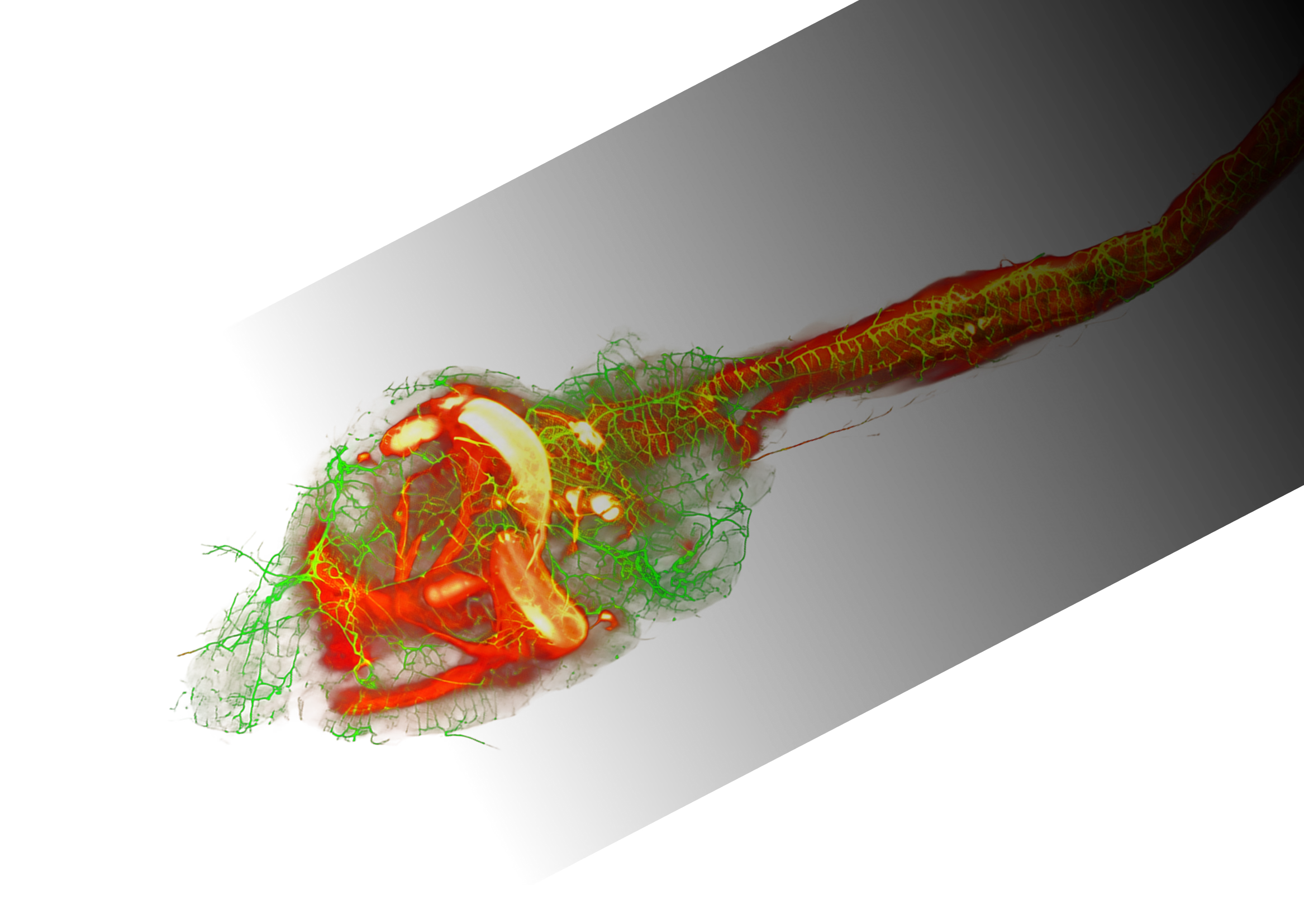
Microscopy visualization of neural tissue

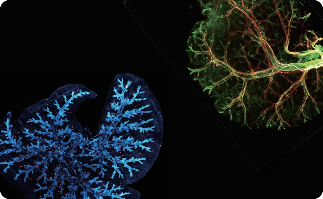
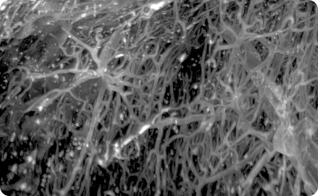
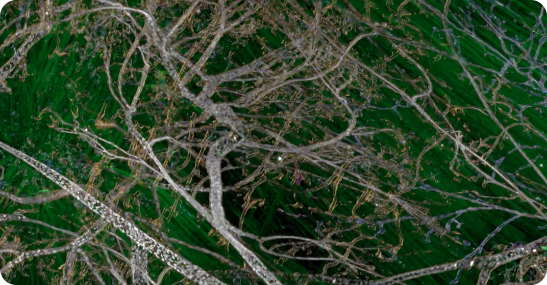
Map vascular networks in 3D to study vessel structure, branching and distribution across tissues

Microscopy visualization of neural tissue

Image larger cleared samples and extended tissue volumes while preserving spatial context and fine detail

End-to-end workflow

Mount
Place the sample in a holder sized to the specimen
Configure
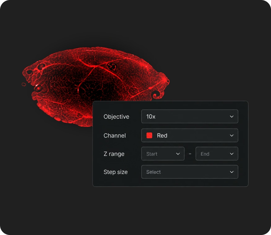
Select the objective and set RI correction
Image
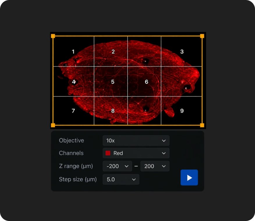
Set positions and tiles, then run automated acquisition
Process
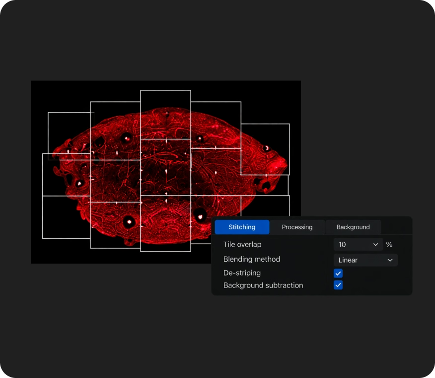
Auto-stitch, fuse and deconvolve in LitScan.
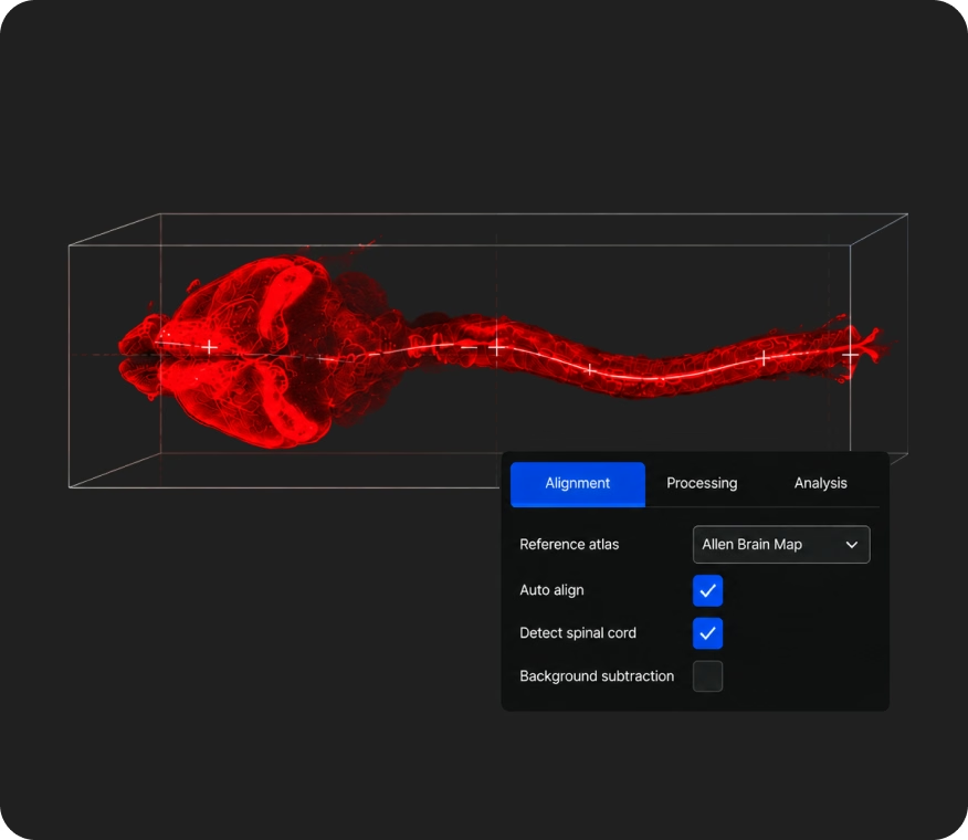
LITScan AiBot analysis visualization
LITScan AiBot
Automated brain and spinal cord alignment to Allen Brain Map. Automates processing and analysis workflows
Analyze
Render in 3D or export to Imaris or Amira

Better 3D imaging comes from technologies designed to work as one

Line Bessel Sheet technology
Long, thin light sheets designed to reduce side-lobe background while preserving axial resolution
Flexible illumination strategies
Illumination approaches adapted to different sample sizes, densities and imaging requirements
RI correction optics
Adjustable refractive-index correction from 1.33 to 1.58 for different clearing media
Integrated processing
Acquisition, stitching, processing and 3D rendering integrated into one workflow

We work alongside you at every stage, from setup to final results

Dr. Anna Keller
”
Research from the lab

Ready to see what your samples can reveal?